

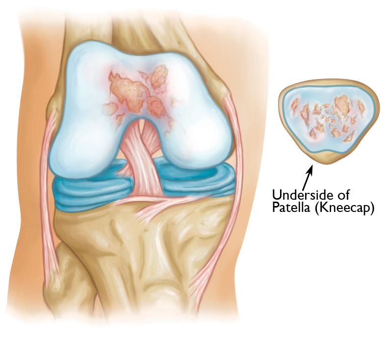
Patellofemoral arthritis affects the underside of the patella (kneecap) and the channel-like groove in the femur (thighbone) that the patella rests in. It causes pain in the front of your knee and can make it difficult to kneel, squat, and climb and descend (go down) stairs.
The patella is a small bone located in front of the knee joint — where the thighbone (femur) and shinbone (tibia) meet. It protects your knee and connects the muscles in the front of your thigh to your tibia.
The patella rests in a groove on top of the femur called the trochlear groove. When you bend and straighten your knee, the patella moves back and forth inside this groove.
A slippery substance called articular cartilage covers the ends of the femur, trochlear groove, and underside of the patella. Articular cartilage helps your bones glide smoothly against each other as you move your leg.
The main symptom of patellofemoral arthritis is pain. Because the patellofemoral joint is in front of the knee, you may have pain in this area. The pain can be present at rest or with no activity at all. Most of the time, however, it is brought on by activities that put pressure on the kneecap, such as kneeling, squatting, climbing and descending stairs, and getting up from a low chair.
In addition, you may experience a crackling sensation called crepitus when you move your knee. Crepitus is sometimes painful and can be loud enough for other people to hear. When the disease is advanced, your kneecap may get stuck, or catch, when you straighten your knee
Patellofemoral arthritis occurs when the articular cartilage along the trochlear groove and on the underside of the patella wears down and becomes inflamed.
When cartilage wears away, it becomes frayed and, when the wear is severe, the underlying bone may become exposed. Moving the bones along this rough surface may be painful.
Dysplasia
Dysplasia occurs when the patella does not fit properly in the trochlear groove of the femur. Because of this, when the knee moves, there are increased stresses on the cartilage. This begins to wear the cartilage down.
Kneecap Fracture
Patellar (kneecap) fractures often damage the articular cartilage that covers and protects the underside of the bone. Even though the broken bone heals, the joint surface may no longer be smooth. There is friction when the patella moves against the joint surface of the femur. Over time, this can lead to arthritis.
When you visit the doctor, they will do a complete medical history and physical examination, as well as order imaging tests.
Your doctor will ask you several questions about your general health, your knee pain, and your ability to function.
It is important for your doctor to determine the exact location of your pain. Patients typically have pain only behind the kneecap, or anterior knee pain. the pain usually occurs during activities that put pressure on the kneecap, such as:
Your doctor will examine the affected knee in various positions to see if there is pain or restricted motion. They will look for creaking or grinding noises (crepitus) that indicate bone-on-bone friction, muscle loss (atrophy), and signs of injury to muscles, tendons, and ligaments.
During the exam, the doctor will:
X-rays. X-rays provide images of dense structures, such as bone. your doctor will order X-rays from several different angles to ensure that your arthritis is limited to the space between the kneecap and the femur, and to assess the overall alignment of your knee.
Magnetic resonance imaging (MRI) scans. MRI scans create better images of the soft tissues in your knee than X-rays do. Your doctor may order an MRI to better evaluate the cartilage in your knee.
Copyright © 2023 Dr. Rahul Bade | All Rights Reserved | Created & Crafted by Itorix Infotech LLP
Need to bounce off ideas for an upcoming project or digital campaign? Looking to transform your business with the implementation of full potential digital marketing?
For any career inquiries, please visit our careers page here.